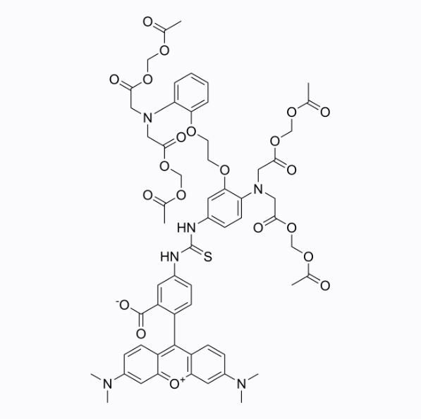
This plan only provides a guide, please modify it to meet your specific needs.
1. Prepare dyeing solution
(1) Prepare stock solution: Prepare AM ester stock solution with a concentration of 1-10mM in high-quality anhydrous DMSO;
Notice:
① Aliquot the unused stock solution and store it in the dark at -20°C or -80°C to avoid repeated freezing and thawing;
② Acetoxymethyl ester (AM) easily absorbs moisture. After taking it out of the refrigerator, please bring it to room temperature in a dry environment before opening it. Please centrifuge briefly before opening to ensure the powder falls to the bottom of the tube.
(2) Prepare working solution: Dilute the stock solution with a suitable buffer (such as serum- and phenol red-free culture medium or PBS) to prepare a working solution with a concentration of 1-10μM.
Notice:
① When preparing the AM ester staining working solution, it is sometimes necessary to add an appropriate amount of 20% Pluronic F-127 solution to the stock solution to enhance the water solubility of the AM probe;
② Pluronic F-127 can prevent AM probes from aggregating in solution and promote better entry of the probe into cells. However, Pluronic F-127 can reduce the stability of the AM probe, so it is only recommended to be added when preparing the working solution. It is not recommended to add the stock solution for long-term storage;
③ Prepare 20% (w/v) Pluronic F-127 stock solution: weigh 100mg Pluronic F-127 powder (Cat. No.: GB30090), add 500μl DMSO, heat at 40-50℃ for 20-30min, and store at room temperature. If crystals precipitate, they can be reheated to dissolve without affecting use;
④ (Optional) GLPBIO provides dissolved Pluronic F-127 (20% Solution in DMSO), product number: GB30091;
⑤ Add an equal volume of 20% Pluronic F-127 solution to the stock solution so that the final working concentration of Pluronic F-127 is approximately 0.02%;
⑥ Please adjust the concentration of the working fluid according to the actual situation, and prepare it now to avoid repeated freezing and thawing.
2. Cell suspension staining
(1) Suspension cells: centrifuge at 4°C and 1000g for 3-5 minutes, discard the supernatant, and wash twice with PBS or other buffer, 5 minutes each time;
(2) Adherent cells: wash twice with PBS or other buffers, add trypsin to digest the cells, and centrifuge at 1000g for 3-5 minutes after digestion is completed;
(3) Add dye working solution to resuspend the cells, and incubate at room temperature or lower than room temperature in the dark for 20min-2h. The optimal incubation time for different cells is different, please explore according to your specific experimental needs;
Note:
① The recommended working concentration of AM ester dyes in most cells is 4-5μM, and the specific concentration needs to be optimized according to experimental requirements. In order to avoid cytotoxicity caused by overloading, it is recommended to use the lowest probe concentration possible based on achieving effective results;
② (Optional) If the cells contain organic anion transporters, you may need to add probenecid (GC16825, Probenecid, 1-2.5mM) or sulfinpyrazone (GC11049, Sulfinpyrazone, 0.1-0.25mM) to the cell culture medium. to reduce the leakage level of the deesterification probe. The stock solution of probenecid or sulfinpyrazone is alkaline, so the pH needs to be readjusted after adding the culture medium;
③ If serum-containing medium is used, serum lactonase will degrade AM, thereby reducing the dye loading effect; while phenol red-containing medium will make the background value slightly higher. It is recommended to wash the cells 2 to 3 times before adding the staining working solution;
④ Lowering the probe loading temperature may reduce the compartmentalization of the probe.
(4) After the incubation, centrifuge at 1000g for 5 minutes to remove the staining solution, add PBS or other buffers and wash 2-3 times to remove residual probes;
(5) Incubate at room temperature for another 30 minutes to ensure complete deesterification of intracellular AM.
3. Cell adhesion staining
(1) Culture adherent cells on sterile coverslips;
(2) Remove the coverslip from the culture medium, suck out the excess culture medium, and place the coverslip in a humid environment;
(3) Add 100 μL of dye working solution from one corner of the coverslip and shake gently to evenly cover all cells with the dye;
(4) Incubate in the dark at room temperature or lower than room temperature for 20min-2h. The optimal incubation time for different cells is different, please explore according to your specific experimental needs;
(5) After the incubation, discard the dye working solution and wash the coverslip 2 to 3 times with PBS or other buffers;
(6) Incubate at room temperature for 30 minutes.
4. Microscope detection: The maximum excitation wavelength and emission wavelength of Calcium Orange AM are 549/576nm respectively.
Precautions:
1) Fluorescent dyes all have quenching problems. Please avoid light as much as possible to slow down fluorescence quenching.
2) For your safety and health, please wear a lab coat and disposable gloves.
References:
[1].Connie M C Lam, et al. Monitoring cytosolic calcium in the dinoflagellate Crypthecodinium cohnii with calcium orange-AM. 2005 Jun;46(6):1021-7. doi: 10.1093/pcp/pci102. Epub 2005 Apr 13.


