


Tylosin/Tylosin A
A macrolide antibiotic, tylosin is active against gram-positive aerobic bacteria and mycoplasmas throughout a wide range of conditions. It is authorized for the treatment of a number of infectious disorders in cattle and poultry, including mastitis, respiratory conditions, and farm animal dysentery. Tylosin, like other macrolide antibiotics, works against bacteria by attaching to the 50S ribosome subunit and preventing the production of new proteins. In this investigation, we used experimental mice models infected with either the avirulent ME49 strain or the virulent RH strain of T. Gondii to assess the anti-T. Gondii capability of tylosin. According to our research, tylosin is effective against T. Gondii both in vitro and in vivo, indicating that it could be a useful treatment for toxoplasmosis in humans.
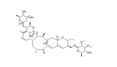
Figure 1 : shows the chemical structure of Tylosin
Tylosin inhibits T. Gondii growth in vitro: In order to examine the effects of Tylosin, cck8 assay was done. Different concentrations of tylosin was given to HFF cells and allow the cells for 48 hours. At 50uM concentration of tylosin, cell proliferation of HFF cells was 99.6 %. At 100uM concentration of tylosin cell proliferation was 88.11%. The 50% inhibitory concentration value was found to be 175.3uM . So the safe concentration of tylosin is found to be ≥ 50 . The assay proved the inhibitory effect of tylosin on parasitic growth(fig.1a).Tylosin was able to inhibit parasite growth, with an EC50 value of 9.759 μM on RH-GFP Strain (Fig. 1b). To better understand the anti-toxoplasma effect of tylosin, Rh strains were treated with 50uM tylosin and 0.1% vehicle. When proliferation rate was examined it was confirm that Tylosin inhibits the activity of extracellular t.gondii tachyzoites(Fig.1c). When tylosin was examined against the intracellular parasites, it proved to inhibit the proliferation of intracellular t.gondii tachyzoites( fig.1d). IFA results confirmed tylosin inhibitory potential against t.gondii infection(fig.1e).
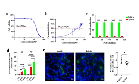
Figure 2:Shows the effect of tylosin treatment on host cell viability and Toxoplasma gondii growth in vitro. A . HFF cells were treated with tylosin at concentrations ranging from 1 to 500 μM for 48 h, and the IC50 value was obtained by CCK8 assays (Glpbio). B . Inhibition of T. gondii type I parasites (RH-GFP) after tylosin treatment. C. Effects of tylosin (50 μM) on the activity of extracellular T. gondii RH tachyzoites. Samples in the control group (Ctrl) were collected at 2 h post infection, and other groups were collected at 48 h or 72 h post infection. Inhibitory effects of tylosin on parasite infection. RH -infected HFFs were treated with tylosin (50 μM) or 0.1% DMSO (vehicle) for 48 h. The cells were stained with DAPI and mouse anti-IMC8 antibody.
Tylosin alters the transcriptome of T. gondii: For the analysis of the effect of tylosin on T. gondii transcriptome, RNA-Seq analysis was performed. Toxoplasma Gondii RH tachyzoites were treated with 50 μM tylosin or 0.1% DMSO (vehicle) for 6 h. A heatmap for the Pearson correlation coefficient between samples was built to investigate the RNA expression patterns. As shown in Fig. 3a, a Pearson correlation matrix clearly highlights the difference between vehicle group and the drug treated group. Principal component analysis (PCA) score plot and unsupervised hierarchical clustering clearly differentiate tylosin-treated groups from vehicle group . Then, a volcano plot was used to analyze differentially expressed (DE) genes. For the better understanding of the roles of these DE genes, Gene Ontology (GO) enrichment analysis was performed. As shown in Fig. 3e, the top 20 most enriched GO terms are listed, and “ribosome biogenesis,” “ribosome” and “gene expression” are the top three most prominent GO terms. Pathway analysis was also performed to determine the affected pathways in parasites following tylosin treatment.
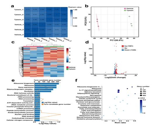
Figure 3: Shows the Incubation with tylosin alters the transcriptome of Toxoplasma gondii RH tachyzoites. A: Heatmap for the Pearson correlation coefficient between samples. B: Principal component analysis (PCA) score scatter plot of Toxoplasma samples . C :Unsupervised hierarchical clustering of R-seq data. D: Volcano plot showing q values (–log10) versus RNA expression ratios between tylosin treatment group and vehicle group (0.1% DMSO). E: Gene Ontology (GO) analysis of DE genes. F : The top 20 significantly enriched KEGG Pathways of the DE genes.
Anti-Toxoplasma activity of tylosin in vivo: KM mice were infected intraperitoneally with 100 T. Gondii RH tachyzoites or 200 ME49 tachyzoites to test the in vivo anti-Toxoplasma activity of tylosin. Mice in mock group were intraperitoneally injected with sterile PBS. Then the infected mice were administered vehicle, tylosin, Sulfadiazine sodium (SDZ) or tylosin+SDZ for 7 consecutive days. At 7 days post infection, vehicle’s group mice showed signs of acute toxoplasmosis, including anorexia, weight loss, edema and messy hair. By Comparing mock group and treatment groups, mice in vehicle group developed severe splenomegaly. After spleen samples staining as shown in Fig. 4b, mice in the vehicle group exhibited a disruption of their splenic architecture with reduced white pulp and increase in the number of megakaryocytes. While on the other hands the spleen index of mice treated with tylosin was significantly reduced compared with mice in the vehicle group as well as the parasite burden in spleen , kidney and lungs was decreased by treatment with the tylosin as shown in the figure 5.
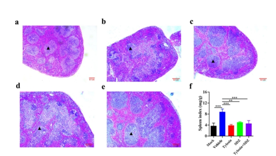
Figure 4: Shows the effect of tylosin treatment on histopathological changes in mouse spleen infected with Toxoplasma gondii RH strain.
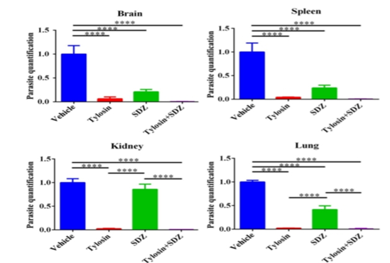
Figure 5 : Shows the effect of tylosin treatment on parasite burden in a. Braun b. Spleen . C. Kidney. d. Lungs .
Tylosin treatment prolonged survival time :Treatment with tylosin reduced the parasite load and resulted in 10% of mice surviving from T. gondii RH strain infection. The combination of tylosin and SDZ exhibited a better synergistic effect and increased the survival rate to 40%. Treatment of mice by tylosin exhibited 40% survival from the ME49 infection. Mice that were treated with the combination of tylosin and SDZ exhibited 70% survival rate.
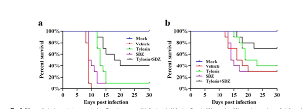
Figure 6 : shows the effects of tylosin on virulent or avirulent Toxoplasmas gondii infections in KM mice. Female KM mice (n=10) were intraperitoneally infected with n=100 RH tachyzoites (a) or n=200 ME49 tachyzoites (b) A negative control group was mock infected with sterile PBS alone. Infected mice were then treated with intraperitoneal injection of tylosin (100 mg/kg/day), oral administration of sulfadiazine sodium (SDZ, 200 mg/Kg/day), tylosin+SDZ, or vehicle. The survival of mice was monitored daily.
In short, Tylosin significantly inhibited the proliferation of T. Gondii in vitro and in vivo. Administration of tylosin significantly extended the survival time and also increased the survival rate of mice infected with T. gondii RH strain or ME49 strain. The combined use f tylosin and SDZ significantly enhanced these effects as compared with each of the monotherapies. Results suggest that tylosin may be the promising agent for the treatment of toxoplasmosis in the future.

 Chemical Structure")


Kommentare