


SP2509 (HCI-2509)
SP2509 also known as HCI-2509 is a small low molecular weight organic compound that can enter into cells and interact with the biological targets .It is used as research tool to study various diseases and biological processes including cancer , inflammation and neurological disorders . It is a selective histone demethylase LSD1 inhibitor is a specific LSD1 antagonist, it can retard tumor cell growth by cell cycle arrest or by inhibiting cell proliferation. To achieve anti tumor effects it can also induce apoptosis in tumor cells. For antiviral functions, SP2509 can hinder herpes simplex virus type 1 (HSV-1) immediate early (IE) gene expression, DNA replication and virus production by inhibiting the LSD1-dependent demethylation of H3K9 on the promoter of HSV-1 IE gene.
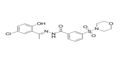
Figure 1: shows the chemical structure of SP2509.
Cytotoxicity of the SP2509 on Vero cells: Researchers first performed CCK8 assay ( Glpbio) in order to determine the effect of different concentrations of SP2509(ranging from 0 to 10 µmol/L: 0, 0.25, 0.5, 1.0, 2.0, 4.0, 8.0, and 10.0 µmol/L) on cell viability. The half-maximal cytopathic concentration (CC50) of SP2509 can be calculated as 4.763µmol/L. Researchers found that SP2509 was not significantly toxic to Vero cells up to 1 µmol/L .The antiviral effects of SP2509 were verified by determining half maximal inhibitory concentration (IC50) on Vero cells as shown in figures.
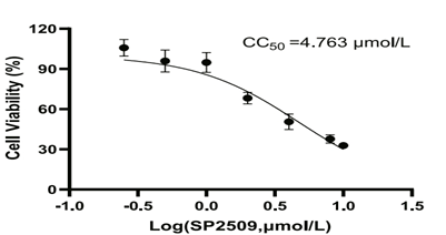
Figure 2: shows maximal inhibitory concentration ( IC50) curves of SP2509.
Antiviral activity of SP2509 on Vero cells : The antiviral effects of SP2509 were confirmed by determining half maximal inhibitory concentration (IC50) on Vero cells. IF method was employed to detect the inhibition rate of SP2509 on PEDV at different concentrations (0.5, 1.0, 2.0, 4.0, 8.0, 10.0 µmol/L), and the IC50 value was determined to be 0.919 µmol/L (Fig. 3A). For the further investigation of the impact of SP2509 on PEDV infection, researchers infected Vero cells with PEDV(0.01MOI) and then exposed the cells to 1 µmol/L SP2509 for the needed time. The virus titer was determined by the collection of virus supernatant . Titer was determined using the TCID50 method to assess the impact of SP2509. The results showed that SP2509 significantly reduced the virus titer compared to the DMSO group (Fig. 3B). Then the researchers conducted RT-qPCR for the analysis of the levels of PEDV ORF3 at different time points. Results showed that the PEDV-ORF3 expression decreased significantly after SP2509 treatment, as shown in Fig. 3C. Next, the researchers collected cells and assessed PEDVN protein expression between the DMSO or SP2509 treated groups. The results of Western blot showed that SP2509 decreased PEDV N protein expression compared to the DMSO group (Fig. 3D). Moreover, the results of immunofluorescence staining indicated that the number of infected cells in the SP2509 treated group was reduced compared to the DMSO treated group (Fig. 3E, F). In short these results demonstrated that a concentration of 1 µmol/L of SP2509 can effectively inhibit PEDV infection in vitro. To test whether the antiviral effect of SP2509 is related to its target LSD1, researchers constructed a Huh-7 cell line capable of knocking down LSD1 and exploring the effect of LSD1 silencing on PEDV infection. The results demonstrated that silencing LSD1 expression significantly suppressed PEDV proliferation as compared with the control group, which is consistent with the results of drug inhibition.
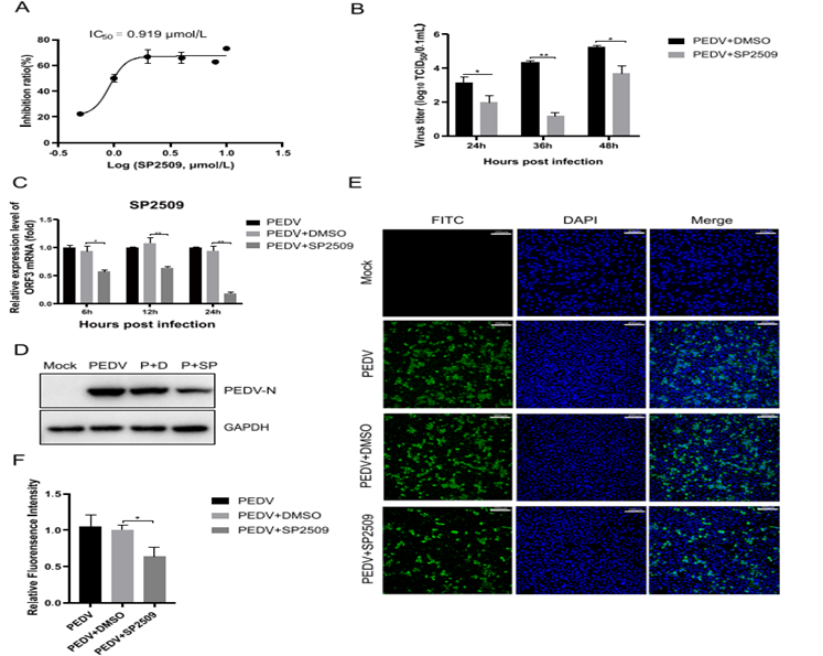
Figure 3: shows how SP2509 inhibits PEDV infection in Vero cells. Vero cells were pretreated with SP2509 for 12 h, followed by infection with PEDV . A: IC50 curves of the SP2509. B : TCID50 analyzed the virus titer. C: RT-qPCR analyzed the Relative RNA expression levels of PEDV ORF3. D : The expression of PEDV N was analyzed by Western blot. E: The inhibitory effect of SP2509 on PEDV N protein was determined by immunofluorescence assay. F: IFA fluorescence quantification results.
SP2509 impaired PEDV internalization and replication instead of attachment or release: For the further exploration of mechanism by which SP2509 inhibits PEDV infection, researchers conducted investigations to identify the specific stage at which SP2509 interferes with PEDV infection (Fig. 4A). For the viral attachment assays, a mixture of 1 µmol/L SP2509 or DMSO and virus (MOI = 0.01) was mixed and added to cells, followed by incubation at 4℃ for 2 h. After washing extensively the treated cells collected to extract RNA to assess the effect of SP2509 on PEDV attachment through RT-qPCR. The observation showed that there is no significant difference in the mRNA level of PEDV-ORF3 , indicating that SP2509 did not affect the attachment of PEDV to the Vero cell (Fig. 4B). To determine the effect of SP2509 on PEDV internalization, Vero cells were infected with PEDV for 2 h at 4 °C. After the removal of the virus inoculum, the supernatant was replaced with 1µmol/L SP2509 or DMSO, and cells were cultured at 37 °C for 3 h. RT-qPCR was then utilized to determine the influence of SP2509 on PEDV-ORF3 expression. According to the results from Fig. 4C, SP2509 impaired PEDV internalization. In the next step , 1µmol/L SP2509 or DMSO was added to the supernatant during the PEDV replication stage. Based on RT-qPCR , PEDV-ORF3 was significantly lower in groups treated with SP2509 (Fig. 4D). This suggested that SP2509 can inhibit the replication phase of PEDV, which was confirmed by immunostaining results in Fig 4 E and F. Finally, Vero cells were continuously infected with PEDV for 24 h. The viral solution was replaced with a maintenance solution, with or without SP2509. After culturing at 37℃ for 4 h, the supernatant was collected, and TCID50 detected the virus titer. The results indicated that the titer of the virus in the supernatant of the SP2509 treated group was not significantly different from that of the DMSO group. This means that SP2509 has not affected the release phase of PEDV (Fig. 4G). Together, the above results implied that SP2509 primarily inhibited PEDV infection by inhibiting virus Internalization and replication.
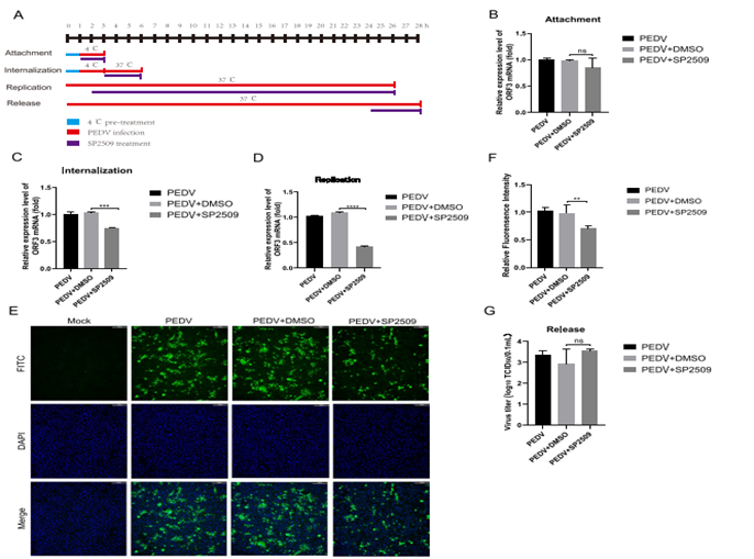
Figure 4: shows The antiviral effect of SP2509 on different infection steps of PEDV. The supernatant was replaced with SP2509 during the virus’s attachment, invasion, replication, and release phases. A : SP2509 treatment schemes. The blue bar represents 4℃ pretreatments, the red bars represent PEDV infection, the purple bars represent SP2509 treatment, and the vertical bars represent the cell collection. B : Virus attachment assay. RT-qPCR detected PEDV-ORF3 mRNA level. C : Virus internalization assay. PEDV-ORF3 mRNA level was detected by RT-qPCR. D: Virus replication assay. PEDV-ORF3 mRNA level was detected by RT-qPCR. E: IFA detection of SP2509 on PEDV replication phase. F: IFA fluorescence quantification results. G : Virus release assay.
In short, African green monkey kidney cells (Vero cells), HEK293T cells and Huh-7 cells were preserved in the Hunan Engineering Technology Research Center of Veterinary Drugs (Hunan Agricultural University, Changsha, China). The cytotoxic effects of SP2509 (GlpBio, ) on Vero cells were assessed using the Cell Counting Kit-8 (CCK-8, Glpbio). Results demonstrated that SP2509 can inhibit PEDV infection during the virus’s internalization and replication stage in vitro. This provided a theoretical basis for the clinical use of SP2509 in anti-PEDV therapy. It inhibits LSD1 activity, depletes colony growth and induces apoptosis and cell death of cultured human acute myeloid leukemia cells.


 Chemical Structure")


Commentaires