


Ademetionine
A ubiquitous metabolite that participates in numerous physiological processes, including transsulfuration, polyamine biosynthesis, and transmethylation is the ademetionine also known as S-adenosyl-L-methionine (AdoMet), SAM or SAMe ( Glpbio ). It serves as the principal methyl donor in the body. Several Studies have highlighted that ademetionine functions as a methyl donor for phosphatidyl ethanolamine, thereby maintains the cell membrane integrity and fluidity. Ademetionine is involved in increasing the concentration of intracellular antioxidants and thus improves cellular oxidative stress by providing polyamines and promoting cell regeneration during metabolism. Recent research on ademetionine has focused on its potential use in treating a variety of conditions, including depression neuropsychiatric disorders , liver disease , osteoarthritis and cancer . Recently researchers assessed the potential benefits of ademetionine in an in vitro oxidative stress-induced MSCs senescence model as well as in an in vivo D-gal induced mouse aging model .
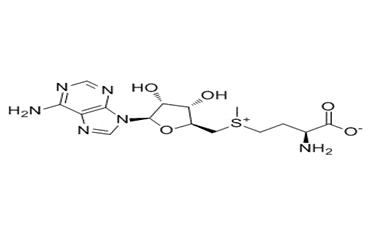
Figure 1: Depicts the chemical structure of ademetionine.
Ademetionine alleviates H2O2-induced senescence of MSCs in vitro: The mesenchymal stem cells (MSCs ) have a spindle-shaped and swirling growth pattern and are obtained from human adipose tissue . Researchers established a senescence model of MSCs by treating them with 100 μM H2O2 for two hours. This concentration of H2O2 was capable of inducing cell senescence instead of apoptosis . In comparison to normal MSCs, Senescent MSCs exhibit higher levels of activity in senescence-associated β-galactosidase as compared to normal MSCs . By treating these cells with varying concentrations of ademetionine (12.5, 25, 50, and 100 μM) for 24 hours, researchers observed a decrease in the percentage of senescence -associated β-galactosidase positive cells and it was concentration dependent. Cell senescence produce high level of reactive oxygen species by dysfunctional mitochondria .Next researchers explored whether exposure to ademetionine reduced ROS levels in MSCs. Following a 24-hour treatment with ademetionine, researchers observed a significant reduction in ROS levels in senescent MSCs and as the concentration of ademetionine was increased ROS levels showed decreasing tendency. By the high concentrations of ademetionine treatment there was a greater increase in SIRT1 protein levels in senescent MSCs. Because of this researchers further selected 50 μM and 100 μM concentrations of ademetionine for subsequent experimental studies. The expression of P53 and P21 was significantly upregulated in senescent MSCs while SIRT1 expression was down regulated but usage of ademetionine mitigated this phenomenon. P53 and P21 are known to regulate cell cycle and cell cycle arrest is also the feature of cell senescence. So the researchers evaluated the cell cycle of MSCs and they found that treatment by ademetionine reduced the number of MSCs in the G1 phase while increased the number of MSCs in the S phase . In short , ademetionine could alleviate H2O2-induced senescence of MSCs in vitro.
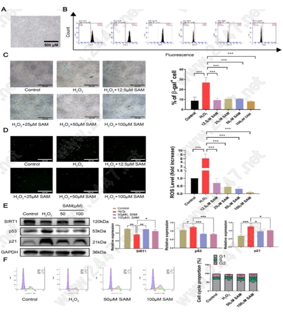
Figure 2: Depicts that how ademetionine alleviates H2O2-induced senescence of MSCs in vitro.
Ademetionine enhances the osteogenic and adipogenic differentiation potential of senescent MSCs in vitro: MSCs have multi-lineage differentiation ability and cell senescence greatly impairs the differentiation potential of MSCs. For the evaluation of the potential effects of ademetionine on MSCs differentiation in vitro, researchers added 50 μM of ademetionine to the differentiation medium of the treatment group and MSCs differentiation was introduced for a period of 0 days, 3 days, 6 days and 9 days. Researchers performed Alizarin red staining for the observation of the calcium nodules generated on the cell surface after 3 days, 6 days and 9 days of inducing osteogenic differentiation. Their analysis revealed a significant reduction in the calcium deposition of the senescent MSCs. Meanwhile, ademetionine treatment significantly increased the calcium deposition and enhanced the osteogenic differentiation potential of the MSCs . In addition, osteoblast genes runx-2, were up regulated as compared with senescent MSCs at day 3, day 6 and day 9 and the RUNX-2 protein expression was also up-regulated after administration ademetionine after 3 days, 6 days and 9 days of inducing osteogenic differentiation . Oxidative stress-senescent MSCs adipogenic differentiation was also promoted by treating with ademetionine and administration of ademetionine also promotes the formation of lipid droplets and increases the expression of early adipocyte-specific genes LPL at day 9 . Furthermore , treatment by ademetionine led to an up-regulation of the LPL protein expression after 6 days and 9 days of inducing adipogenic differentiation, thereby promoting adipogenic differentiation in the MSCs .
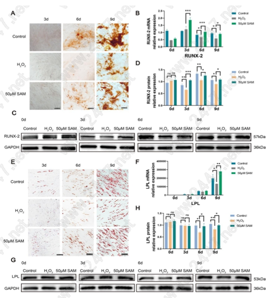
Figure 3: Depicts that how the Ademetionine enhances the osteogenic and adipogenic differentiation potential of senescent MSCs in vitro.
Ademetionine exerts anti-aging roles in vivo in D-gal induced mouse aging model:Researchers constructed a premature aging mouse model for the further investigation of the potential anti-aging effects of ademetionine in vivo .The animal experiments were consisted of three stages. In the first stage (from the second to the sixth week) mice were subcutaneously injected with D-galactose (150 mg/kg/d) in the neck and back. In the second stage (from the sixth to the eighth week), D-gal injection was continued and ademetionine was administered daily by gavage. In the third stage (from the eighth to the ninth week), mice were only treated with ademetionine (30Mg/kg/d) by gavage every day. Researchers performed the open field test and the rota rod test in order to measure the overall anti-aging effects of ademetionine on mice . The behavioral tests depicted that exercise ability and endurance were significantly improved in the ademetionine group compared to the D-gal group . A question may arise What are the effects of SAM on the major organs such as the heart, liver and kidney? Answers is that the ademetionine also reduced the expression of P16INK4a in mouse liver tissue, as well as reduced the expression of P53 in mouse kidney tissue and heart tissue . Immunohistochemical staining for P53 in the heart, liver and kidney tissues of mice showed that P53 was positive mainly in cardiomyocytes, pericentral veins of hepatic lobules and glomeruli, which could be alleviated by SAM treatment . These results highlight the beneficial anti-aging roles of ademetionine on main organs such as heart, liver and kidney. Finally, to investigate the anti-aging mechanism of ademetionine in vivo, researchers examined the activity of serum superoxide dismutase (SOD) and glutathione peroxidase (GPX) as well as the content of malondialdehyde (MDA).
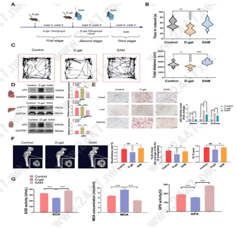
Figure 4: Shows how the Ademetionine exerts anti-aging roles in vivo in D-gal induced mouse aging model.
PI3K/AKT pathway was activated by ademetionine to mitigate the oxidative stress-induced senescence of MSCs: H2O2 treatment reduced the levels of PI3K, p-PI3K, AKT, and p-AKT while administration of ademetionine reversed their levels . This suggests that ademetionine relieves oxidative stress-induced senescence of MSCs by activating the PI3K/AKT pathway. To verify the role of PI3K/AKT in ademetionine’s anti-senescence effects, LY294002, a broad-spectrum PI3K inhibitor, was used to pre-treat MSCs. The usage of LY294002 further inhibited PI3K/AKT pathway in combination with H2O2. Surprisingly, ademetionine up-regulated the levels of p-PI3K, p-AKT and SIRT1, while down-regulating P53 level. Thus, ademetionine exerted its anti- senescence role by activating the PI3K/AKT pathway .Researchers also examined the role of PI3K in SAM-induced cell protection. SAM treatment decreased the ROS levels in senescent MSC while this reduction was reversed by the addition of LY294002 . Furthermore, to demonstrate the effect of LY294002 on ademetionine’s promotion of MSC differentiation, researchers induced differentiation of MSCs for 12 days using an induction solution containing SAM with or without LY294002. The results depicted that LY294002 reduced the calcium deposition and lipid droplet formation decreased the gene expression of runx-2 and PPARy-γ at the sixth day and diminished the protein levels of PPAR-γ and RUNX-2 . These results show that the anti-senescence effect of SAM on MSCs can be inhibited by the
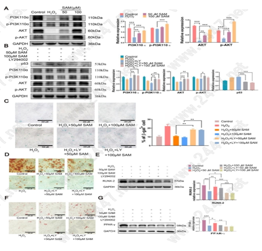
Figure 5 : ( A ) Representative images of western blot and the quantification analysis of PI3K/AKT pathway related proteins. (B) Representative images of western blot and quantitative analysis of pathway core gene expression and senescence related protein expression. © Representative images of β-galactosidase staining. Scale bar, 200 μm. (D) Representative images of Alizarin red staining. Scale bar, 200 μm. (F) Representative images of oil red O staining. Scale bar, 200 μm. (E and G) Representative images of western blot analysis of PPAR-γ and RUNX-2.
In short, these findings highlight that the PI3K/AKT/FOXO3a axis in MSCs could play a crucial role in MSCs senescence and suggest that ademetionine may be a potential therapeutic drug for MSCs senescence and related diseases. These findings may open avenues for developing innovative therapeutic strategies to address MSC senescence-related issues.


コメント