D-Luciferin (sodium salt) |
| カタログ番号GC43497 |
D-ルシフェリン (D-(-)-ルシフェリン) ナトリウムは、生物発光昆虫の発光を触媒するルシフェラーゼの基質です。
Products are for research use only. Not for human use. We do not sell to patients.

Cas No.: 103404-75-7
Sample solution is provided at 25 µL, 10mM.
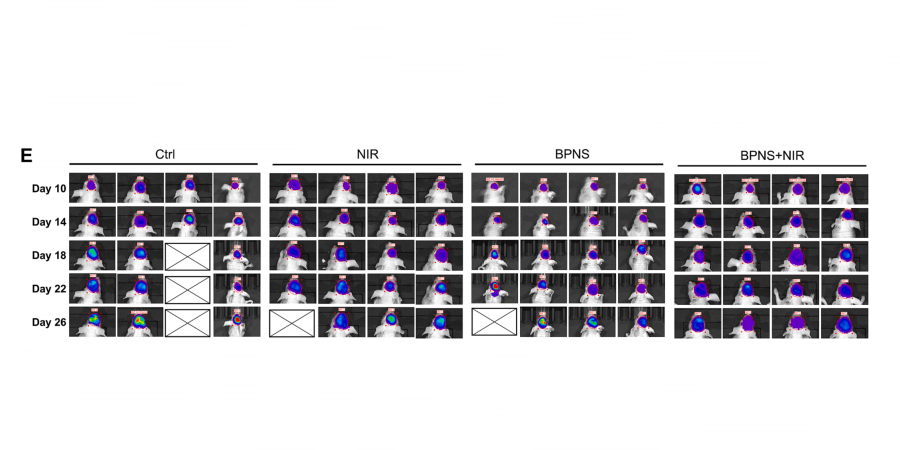
D-luciferin is the natural substrate of firefly luciferase. In the presence of magnesium ions, luciferase catalyzes the reaction of luciferin with ATP, which is then oxidized to form a dioxetane structure that emits yellow-green light [1]. When the substrate luciferin is in excess, the 560 nm chemiluminescence generated by the Luciferin-luciferase luminescent reaction reaches its peak within a few seconds, and the light output is proportional to the luciferase concentration. Chemiluminescent techniques are virtually background-free, making the luciferase reporter an ideal tool for detecting low-level gene expression. 0.02 pg of luciferase can be reliably measured in a standard fluorescence counter. D-luciferin is a commonly used reporter gene for ATP detection, cell viability assay, reporter gene detection, active molecular screening and bacterial counting. D-luciferin is widely used in live animal imaging. Cells expressing the luciferase gene were transplanted into research animals and injected with D-luciferin to be able to detect changes in brightness by bioluminescence imaging (BLI)[2].
D-Luciferin has three product forms, D-Luciferin (D-Luciferin, free acid), D-Luciferin potassium salt (D-Luciferin, potassium salt) and D-Luciferin sodium salt (D-Luciferin, sodium salt ). The potassium and sodium salt forms of D-fluorescein are the most versatile because they are both readily soluble in water. Potassium salt is also the main form used in live animal testing.
References:
[1]. Giuseppe Meroni, et al. D-Luciferin, derivatives and analogues: synthesis and in vitro/in vivo luciferase-catalyzed bioluminescent activity. ARKIVOC 2009 (i) 265-288.
[2]. Sangyub Kim, et al. Optimizing live-animal bioluminescence imaging prediction of tumor burden in human prostate cancer xenograft models in SCID-NSG mice.2019 Jun;79(9):949-960. doi: 10.1002/pros.23802. Epub 2019 Apr 8.
Ⅰ. Matters needing attention
1. D-luciferin is easily soluble in aqueous buffer solution (pH 6.1-6.5), and the solubility can reach up to 100 mM. For example: Use sterile D-PBS (without Ca2+ and Mg2+) to prepare D-luciferin potassium salt solution (15mg/ml), and filter it with a 0.22µm filter membrane (protect from light). D-luciferin solution is recommended to be prepared and used immediately. It can also be frozen and stored at -20°C or -80°C after aliquoting to avoid repeated freezing and thawing. Melt at 4°C before use, and equilibrate to room temperature before experiment (protect from light)
2. If used to detect ATP, please wear gloves and use ATP-free autoclaved water, reagents and containers to minimize all possible sources of ATP contamination.
Ⅱ. Experimental method:
The following scheme is an example sodium salt preparation of potassium and potassium. It is suitable for most cell types and in vivo animal use.
This protocol provides a guide only and should be modified according to your specific needs.
1. In vitro (intracellular) fluorescence imaging
(1) Seed cells stably expressing luciferase in a 12-well plate (2×10³/well).
(2) Use sterile water to prepare 100 mM fluorescein stock solution, and after fully dissolved, use a 0.22 µm filter membrane to filter and sterilize (protect from light).
(3) Use pre-warmed medium to dilute the stock solution to a working solution with a concentration of 0.5-1 mM
(4) Aspirate the medium from the cultured cells, add fluorescein working solution to the cells, and incubate the cells at 37°C for 5-10 minutes before imaging[1].
(5) Use a series of filters (520- 800nm) for image acquisition
2. In vivo fluorescence imaging
(1) Use sterile D-PBS (without Ca2+ and Mg2+) to prepare D-luciferin potassium salt stock solution (15mg/ml), and after fully dissolved, use a 0.22µm filter membrane to filter and sterilize (protect from light).
(2) Inject animals intraperitoneally 10-15 minutes before imaging, at a dosage of 75-150mg/Kg [2] [3].
(3) Fluorescent imaging of experimental animals using bioluminescent imaging
Note: Fluorescein kinetic studies should be performed for each animal model to determine peak signal time.
References:
[1]. Giuseppe Meroni, et al. D-Luciferin, derivatives and analogues: synthesis and in vitro/in vivo luciferase-catalyzed bioluminescent activity. ARKIVOC 2009 (i) 265-288.
[2]. Wentian Zhang,et. Dual inhibition of HDAC and tyrosine kinase signaling pathways with CUDC-907 attenuates TGFβ1 induced lung and tumor fibrosis. 2020 Sep 17;11(9):765. doi: 10.1038/s41419-020-02916-w.
[3]. Senlin Li,et. Concurrent silencing of TBCE and drug delivery to overcome platinum-based resistance in liver cancer. 2023 Mar;13(3):967-981. doi: 10.1016/j.apsb.2022.03.003. Epub 2022 Mar 12.
| Cas No. | 103404-75-7 | SDF | |
| Canonical SMILES | O=C([O-])[C@H]1CSC(C(S2)=NC3=C2C=C(O)C=C3)=N1.[Na+] | ||
| Formula | C11H7N2O3S2•Na | M.Wt | 302.3 |
| 溶解度 | DMSO : 100 mg/mL (330.80 mM); Water : 80 mg/mL (264.63 mM) | Storage | Store at -20°C, protect from light |
| General tips | Please select the appropriate solvent to prepare the stock solution according to the
solubility of the product in different solvents; once the solution is prepared, please store it in
separate packages to avoid product failure caused by repeated freezing and thawing.Storage method
and period of the stock solution: When stored at -80°C, please use it within 6 months; when stored
at -20°C, please use it within 1 month. To increase solubility, heat the tube to 37°C and then oscillate in an ultrasonic bath for some time. |
||
| Shipping Condition | Evaluation sample solution: shipped with blue ice. All other sizes available: with RT, or with Blue Ice upon request. | ||
| Prepare stock solution | |||

|
1 mg | 5 mg | 10 mg |
| 1 mM | 3.308 mL | 16.5399 mL | 33.0797 mL |
| 5 mM | 0.6616 mL | 3.308 mL | 6.6159 mL |
| 10 mM | 0.3308 mL | 1.654 mL | 3.308 mL |
Step 1: Enter information below (Recommended: An additional animal making an allowance for loss during the experiment)
 g
g
 μL
μL

Step 2: Enter the in vivo formulation (This is only the calculator, not formulation. Please contact us first if there is no in vivo formulation at the solubility Section.)
Calculation results:
Working concentration: mg/ml;
Method for preparing DMSO master liquid: mg drug pre-dissolved in μL DMSO ( Master liquid concentration mg/mL, Please contact us first if the concentration exceeds the DMSO solubility of the batch of drug. )
Method for preparing in vivo formulation: Take μL DMSO master liquid, next addμL PEG300, mix and clarify, next addμL Tween 80, mix and clarify, next add μL ddH2O, mix and clarify.
Method for preparing in vivo formulation: Take μL DMSO master liquid, next add μL Corn oil, mix and clarify.
Note: 1. Please make sure the liquid is clear before adding the next solvent.
2. Be sure to add the solvent(s) in order. You must ensure that the solution obtained, in the previous addition, is a clear solution before proceeding to add the next solvent. Physical methods such as vortex, ultrasound or hot water bath can be used to aid dissolving.
3. All of the above co-solvents are available for purchase on the GlpBio website.
Quality Control & SDS
- View current batch:
- Purity: >98.00%
- COA (Certificate Of Analysis)
- SDS (Safety Data Sheet)
- Datasheet
Average Rating: 5 (Based on Reviews and 8 reference(s) in Google Scholar.)
GLPBIO products are for RESEARCH USE ONLY. Please make sure your review or question is research based.
Required fields are marked with *


