


Px -478
Chemically, the full name of PX 478 is S-2-amino3-[4’-N, N,-bis(chloroethyl)amino]phenyl propionic acid N-oxide. It is a small molecule, i.e., having low atomicity and molecular weight. The 2D view of the molecular structure of the PX 478 compound is schematically depicted in Figure 1.
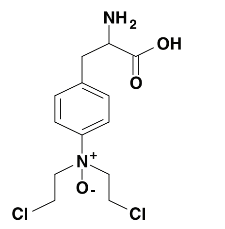
Figure 1 The 2D view of the Molecular Structure of the PX 478 compound
PX 478 inhibits Hypoxia Inducible Factor 1α (HIF-1α). The domains of HIF-1α are shown in Figure 2.
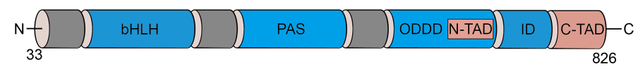
Figure 2 A cartoon diagram of the Hypoxia Inducible Factor 1α (HIF-1α) protein showing its labelled distinct functional domains. basic helix-loop-helix (bHLH) domain that bind with the DNA double helix, PAS domain whose function is in different protein/ protein interactions and dimerization process, oxygen-dependent degradation domain (ODDD), N-terminal trans-activation domain (N-TAD), inhibitory domain (ID) and C-terminal trans-activation domain (C-TAD).
Hypoxia Inducible Factor 1α (HIF-1α) is a transcription factor; thus, after being expressed it gets translocated in the nucleus where it promotes the transcription of its target genes.
Hypoxia Inducible Factor 1α (HIF-1α) is a protein that is expressed especially in those cells which are marked by hypoxia, a stress condition in which there is a low availability of the molecular oxygen to the cells. A perfect example of such cells are the tumor cells. In the tumor cells, abnormally upregulated expression of Hypoxia Inducible Factor 1α (HIF-1α) causes an increase in the vascularization; resultantly, also an increase in the energy metabolism. This is because high vascularization eventually increases the available molecular oxygen. This observation is clinically significant and also indicates poor prognosis that meaning cancer is aggressively propagating. This is true for a number of cancers/ neoplasms, for example, oropharyngeal cancer, esophageal cancer, lung cancer, breast cancer, ovarian cancer, cervical cancer, etc. Therefore, inhibiting Hypoxia Inducible Factor 1α (HIF-1α) via the introduction of small molecule inhibitor, like PX 478 is a novel strategy to treat a broad spectrum of neoplasms.
Besides acting as anti tumor, PX 478 also reduces atherosclerosis. Atherosclerosis is a chronic inflammatory disease of the blood arteries in which the Atherosclerotic lesions are marked by severe hypoxia, a stress condition. In atherosclerosis, hypoxia triggers inflammation, formation and enlargement of plaque, along with the modelling of the arteries. Overall, the clinically significant effects of PX 478 treatment on the atherosclerosis models of mice are summarized in Figure 3.
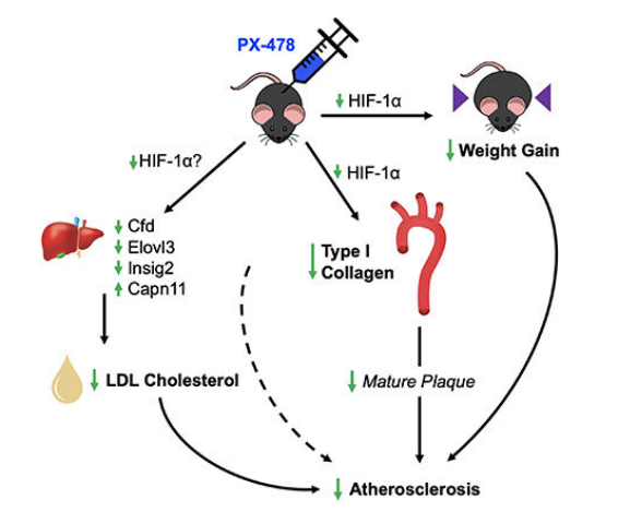
Figure 3 The PX 478 treatment ultimately lessens the atherosclerosis via three main mechanisms, first by lowering the amount of Low Density Lipids (LDL) in the blood plasma, second by lowering the Type 1 Collagen, and third by lowering the weight.
Under the normal physiology, i.e., in the adequate availability of molecular oxygen or in other words in the absence of hypoxia, prolyl hydroxylase domain (PHD) proteins add hydroxyl group to the proline residues of the Hypoxia Inducible Factor 1α (HIF-1α) protein; leading to the binding of von Hippel-Lindau protein (pVHL) with the Hypoxia Inducible Factor 1α (HIF-1α) protein, eventually Hypoxia Inducible Factor 1α (HIF-1α) protein is ubiquitinated and destined to be biochemically degraded via the activity of the proteasomal enzymes. Therefore, under normal physiological condition Hypoxia Inducible Factor 1α (HIF-1α) protein is not present as it is continuously being degraded. While on the other hand, under the hypoxia condition, HIF-1α protein does not get hydroxylated by the activity of prolyl hydroxylase domain (PHD) proteins as prolyl hydroxylase domain (PHD) proteins are itself downregulated. Therefore, the expression of HIF-1α protein is upregulated in hypoxia as shown in Figure 4.
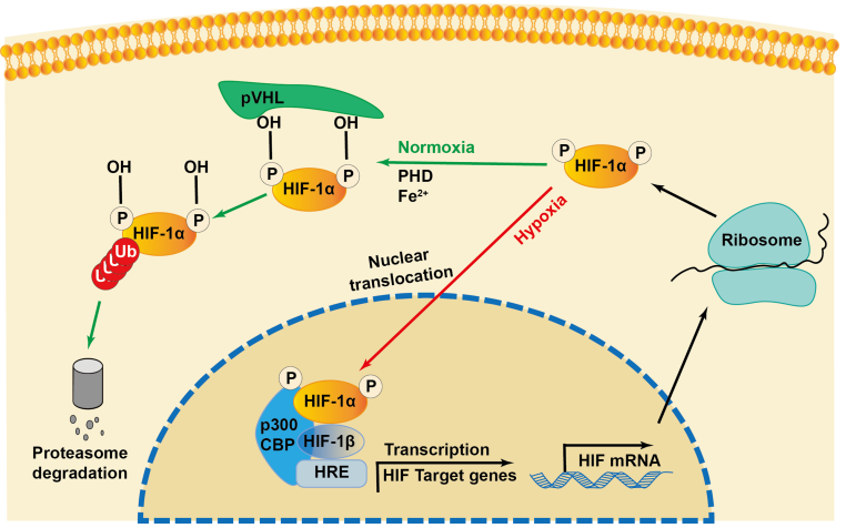
Figure 4 The down regulation of Hypoxia Inducible Factor 1α (HIF-1α) protein under the normal physiology which is marked by normoxia as well as the up regulation of the same under abnormal pathology which is marked by hypoxia
PX 478 inhibits the protein expression of Hypoxia Inducible Factor 1α (HIF-1α) in a dose dependent manner and also, complete inhibition of Hypoxia Inducible Factor 1α (HIF-1α) takes place at 100 mM concentration. This observation is evident from the gel image from the western blotting as shown in Figure 5 and is also graphically presented in Figure 6.

Figure 5 The PX 478 treatment inhibits the protein expression of Hypoxia Inducible Factor 1α (HIF-1α) in a dose dependent manner
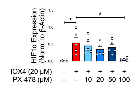
Figure 6 The PX 478 treatment inhibits the protein expression of Hypoxia Inducible Factor 1α (HIF-1α) in a dose dependent manner
The PX 478 treatment also prevents the RNA transcription of the target genes of Hypoxia Inducible Factor 1α (HIF-1α) namely EDN1, GLUT1, and LOX. In other words, decreased levels of Hypoxia Inducible Factor 1α (HIF-1α) protein causes a decrement in the transactivation activity associated with Hypoxia Inducible Factor 1α (HIF-1α). This observation is evident from the results of qPCR that are graphically presented in Figure 7. Other target genes like VEGF and GLUT3 are also found to be downregulated in other type of cell lines (data not shown).
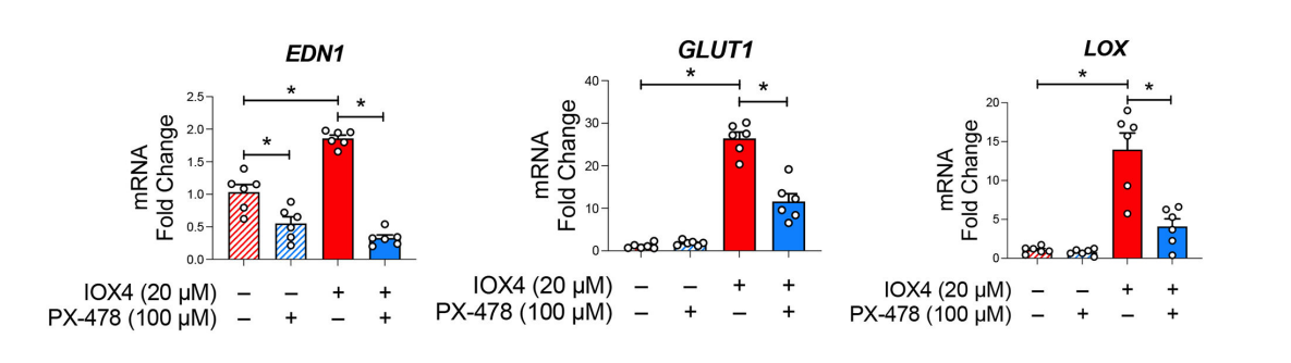
Figure 7 The PX 478 treatment also prevents the RNA transcriptions of the target genes of Hypoxia Inducible Factor 1α (HIF-1α). It was previously mentioned that Hypoxia Inducible Factor 1α (HIF-1α) is a transcription factor, i.e., it upregulates the RNA transcription of certain genes.
In the murine model of atherosclerosis, the anti-atherogenic properties of the PX 478 compound has been confirmed against the saline solution. These observations are given in Figure 8 (in the form of Oil-Red O stained images of the aortic tree), Figure 9 (graphically presented), Figure 10 (decreased plaque area), Figure 11 (decreased lipid lesion area), Figure 12 (decreased mucin area) and Figure 13 (decreased Type 1 Collagen and Total Collagen) using various histological stains.
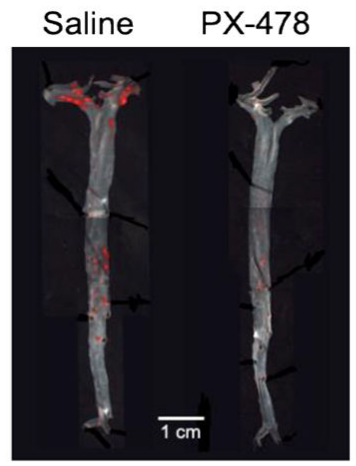
Figure 8 The Oil-Red O stained images of the aortic trees of the two murine model of atherosclerosis. A mouse was treated with saline as shown in left image; while on the other hand, another mouse was given treatment with PX 478 as shown in right image. The red color indicates plaque burden; and it is surely less in the PX 478 treated mouse model of atherosclerosis
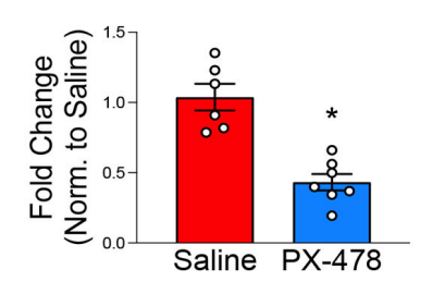
Figure 9 The plaque burden is significantly higher in the saline treated mouse than the PX 478 treated mouse; this indicates that the PX 478 treatment prevents the development of arteriosclerosis. This graph is generated by the quantification of the same Oil-Red O stained images which are presented in Figure 6 via MATLAB software.
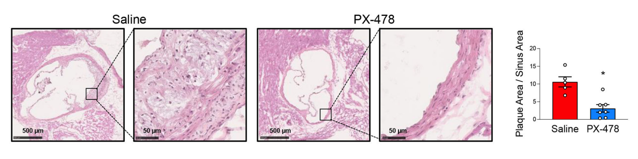
Figure 10 The PX 478 treatment significantly decreases the size of plaque in terms of area as compared to saline treatment as an experimental control
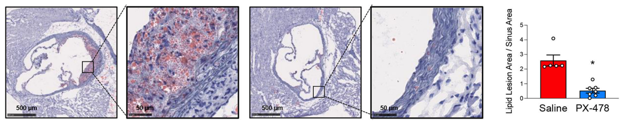
Figure 11 The PX 478 treatment significantly decreases size of the lipid lesion in terms of area as compared to saline treatment as an experimental control. Left pair is saline treated and right pair is PX 478 treated.
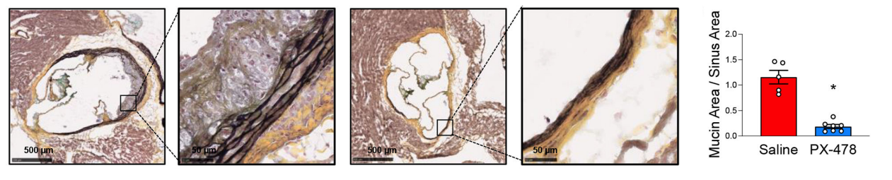
Figure 12 The PX 478 treatment significantly decreases size of the Mucin in terms of area as compared to saline treatment as an experimental control. Left pair is saline treated and right pair is PX 478 treated.
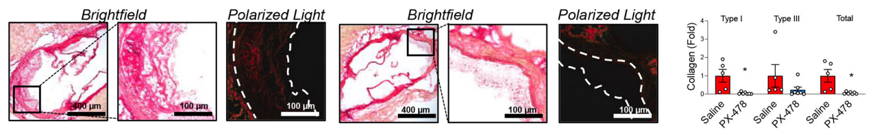
Figure 13 The PX 478 treatment significantly decreases in total collagen and mature Type 1 collagen as compared to saline treatment as an experimental control. Three pictures on the left are saline treated and three pictures on the right are PX 478 treated.


Comments