


SBFI-26
SBFI-26 is a fatty acid binding protein inhibitor and it binds to anandamide transporters FABP5 and FABP7 at two different sites. Traditional Chinese medicine is Incarvillea Sinensis and the SBFI-26 is the key active component of this medicine. When applied to mouse models this compound shows efficient anti- tumor roles. By acting as anti-tumor agent it is used to treat castration-resistant prostate cancer (CRPC). It interferes with the FABP5-PPARγ signaling pathway when it is at the initial stage of the signal transduction .It binds competitively to FABP5 to inhibit cellular fatty acid uptake. SBFI-26 also produces anti-nociceptive and anti inflammatory effects.
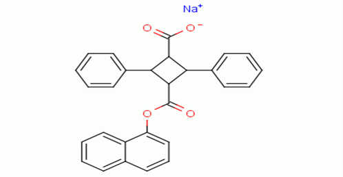
Figure 1 : Shows the chemical structure of SBFI-26
To evaluate the mechanism of cytotoxicity of SBFI-26 on breast cancer cell lines , MCF-7, MDA-MB-468 and MDA-MB-231 cells were treated with varying concentrations of SBFI-26 for different durations (12, 24, or 48 h). By performing a CCK-8 kit assay cell viability was evaluated .As depicted in Figures 2B–D, The result demonstrates that SBFI-26 has significantly reduced the proliferation of MDA-MB-468 and MDA-MB-231 cell lines in a both concentration-dependent and time-dependent manner. The inhibitory effects of SBFI-26 on cell proliferation were significantly different from those of the control group after 12 h of treatment across all three cell lines. After 24 h of the treatment, only MDA-MB-468 and MDA-MB-231 cells showed marked differences in inhibition as compared to the control group at high doses (125 and 150 μM). After 48 h of treatment, SBFI-26 showed marked inhibitory activity on MDA-MB-468 and MDA-MB-231 cells as compared to the control group. However, MCF-7 has no any significant time dependence, the inhibition by SBFI-26 on MCF-7 cells was observed only at doses greater than 100 μM and the inhibition rate could reach 50%. Notably, SBFI-26 demonstrated superb efficacy against MDA-MB-231 cells. These cells belong to the TNBC subtype and exhibit high expression levels of FABP5 protein that meke them more sensitive to SBFI-26. Therefore, subsequent studies were conducted using the MDA-MB-231 cell line.
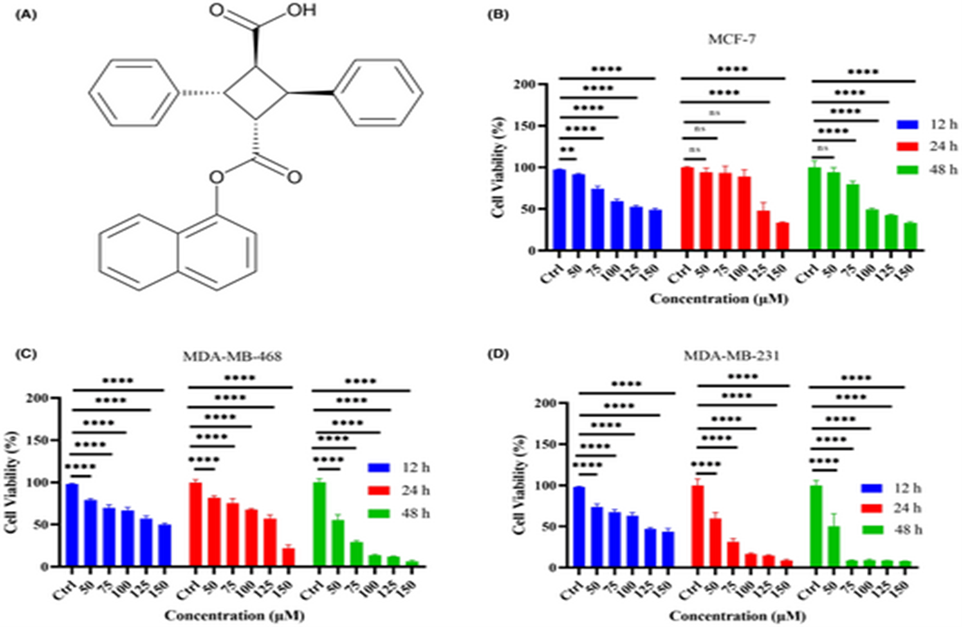 Figure 2: shoes SBFI26 suppresses breast cancer cell growth and induces cell death by suppressing cell growth and proliferation.
(A) The chemical structure of SBFI26. (B–D) Show the Cell viability of MCF-7, MDA-MB-468, and MDA-MB-231 cells and was measured by CCK8 assay after treatment with known concentration of SBFI26 (50, 75, 100,125,150 μM) at 12, 24, 48 h. Statistical analysis was carried out between the SBFI-26-treated group and the untreated group. Data were presented as Mean ± SD. Data were analyzed by the use of one-way ANOVA, with p < 0.05, p < 0.01, p < 0.001, p < 0.0001; ns, not significant.
Castration resistant-prostate cancer is largely dormant to feather by therapy and hence the outlook for patients is hard and grim. Here is the approach to attach the recently discovered Achilles heel. The experimental treatment that is established in the study is based on the recent discovery that is the FABP5-PPARγ-VEGF signaling axis, rather than the androgen receptor pathway, FABP5-PPARγ-VEGF played a dominant role in promoting the malignant progression of castration resistant prostate cancer cells. Treatments that have been established in mice are done by suppressing the biological activity of FABP5 by using a chemical inhibitor SBFI-26. The inhibitor markedly suppressed the proliferation, migration, invasiveness and colony formation of PC3-M cells in vitro. It also caused a highly noticeable suppression of both the metastasis and the primary tumors developed from cancer cells implanted orthotopically into the prostate glands of the mice. The question arises how the inhibitor SBFI-26 interferes with the FABP5-PPARγ signaling pathway? It interferes with the initial stage of signal transduction by binding competitively to FABP5 to inhibit cellular fatty acid uptake. Reduced fatty acid uptake avoids the fatty acid stimulation of PPARγ and prevents it to activate the downstream regulated cancer-promoting genes. This is entirely new experimental approach to treat castration-resistant prostate cancer is completely different from current treatments that are based on androgen-blockade therapy.
Cytotoxicity tests demonstrated that treatment with SBFI-26 significantly suppressed viability of PC3-M cells in a concentration dependent pattern. Suppression was Maximum at concentration of 100 μM for SBFI-26, further increase in doses did not produced any significant suppression. When treated with this optimal dose cell numbers were reduced by 26% (student’s t test, P < 0.001) in figure 3A. When tested using a MTT assay, 100 μM Concentrated SBFI-26 a marked increase in the proliferation rate of PC3-M cells by 17 times was observed (student’s t test, P < 0.001) in figure 3B. When tested in a cell migration assay in figure 3C treatments with 100 μM SBFI-26 . It produced only 19% reduction in wound size in 24h. This treatment significantly suppressed the migration rates of PC3-M cells (student’s t test, P < 0.001), leading only to small changes in wound gaps for the treated group compared to an almost gap closure (94%) for the control in figure 3D. When this was tested in an invasion assay, then the mean number of invaded cells from the control and the PC3-M cells treated with SBFI-26 were 22 ± 3 and 1 ± 1, respectively, representing a highly significant suppression of invasion by 95.5% (student’s t test, P < 0.001)in figure 3E. It was further tests in soft agar and it showed that the number of colonies formed after two weeks by control PC3-M cells and PC3-M cells treated with SBFI-26 were 124 ± 18 and 0 respectively , representing a highly significant inhibition by 100% (student’s t test, P < 0.001) in figure 3F.
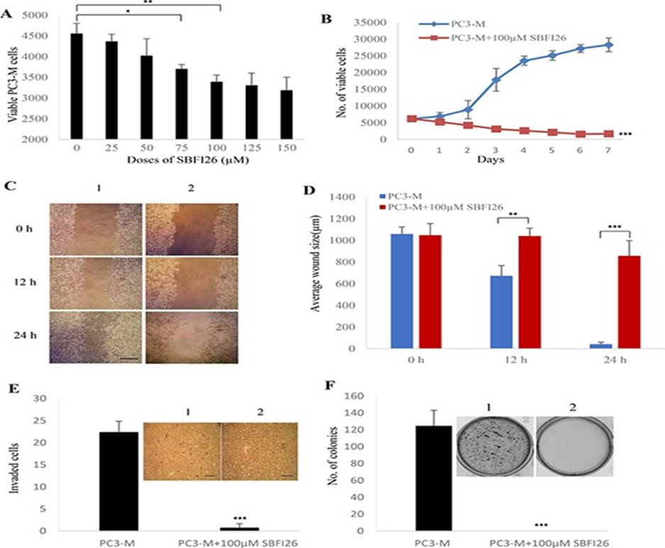
Figure 3: shows inhibitory effect of SBFI26 on malignant characteristics of PC3-M cells.
Determination of the optimal inhibitory concentration of SBFI26 :At this concentration the maximum suppression of cell growth is achieved. MTT assay was performed to measure the viable PC3-M cell numbers of the control (untreated) and those treated with different concentrations of SBFI26 for 24 h. (B) Demonstrates Inhibitory effect of 100 μM SBFI26 on proliferation of PC3-M cells over the 7day experimental period. (C) Representative photos of the wound healing assay. PC3-M cells were grown in 6-well plates to form a monolayer. Scratches were made using 1 mL sterile pipette tip. Measured the cell migration capacity by the reduction in wound size in control (1) and in cultures treated with 100 μM SBFI26 (2) observed at 0, 12 and 24 hours after the treatment. The scale bar is at 250 μm. (D) Average wound sizes (μm) of the control PC3-M and cultures treated with 100 μM SBFI-26 observed at 0, 12 and 24 hours after treatment. Byy measuring image of the wound space data was collected and analyzed by Image software (National Institutes of Health). (E) Number of invading cells from the control PC3-M cells (1) and cultures treated with 100 μM SBFI26 (2) for 24 h after different treatments. Results (mean ± SE) were obtained from three separate measurements. Scale bar is 250 μm. (F) Colonies produced by the control PC3-M cells (1) and cultures treated with 100 μM SBFI26 (2) in soft agar 2 weeks after the different treatments. Results (mean ± SE) were obtained from three separate plates in each treatment. The inserted picture was a representative plate from each of the 3 treatments. All in vitro results were subjected to 2-tailed unpaired Student’s t test and P < 0.05; P < 0.001; P < 0.0001.
Over the past many years, SBFI-26 and related compounds have been actively developed as potential analgesics that target the intracellular anandamide transporters FABP5 and FABP77. High-resolution structures of SBFI-26 in complex with the proteins. These complexes would aid our medicinal chemistry efforts to design more potent inhibitors based on the SBFI-26 scaffold and our research manifested that in FABP5, inhibitor SBFI-26 down regulates PC3-M cells to inhibit cancer development in these cells and also breast cancer cell growth and that induce death in these cells.

 Chemical Structure")


Comments