Geneticin
Other names of Geneticin include G418 and G418 Sulphate. Pharmaceutically, Geneticin is an antibiotic belonging to the category of aminoglycoside. Aminoglycosides are the compounds having the amino group bounded to the sugar moiety. Its antibiotic effect, specifically the antiviral effect has been confirmed against bovine viral diarrhea virus (BVDV), dengue virus (DENV).
Geneticin is a small molecule. The molecular structure of the Geneticin compound is presented in Figure 1.
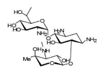
Figure 1 The Molecular Structure of the Geneticin compound
In BHK cells, the Geneticin treatment prevents cytopathic effects (CPE) and thereby improves the Cell Viability (percentage of survival) specifically in the dengue virus Type 2 (DENV2) infected cells, in a dose dependent manner. In addition, it also reduces viral yield, plaque formation, expression of Viral E protein and synthesis of viral RNA. These observations are graphically presented in Figure 2, Figure 3, Figure 4, Figure 5, and Figure 6 respectively. On top of that, Geneticin is specific in its inhibitory action. Yet, there is not known molecular basis for this antiviral effect .
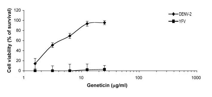
Figure 2 The Geneticin treatment improves the Cell Viability (percentage of survival) only in the dengue virus Type 2 (DENV2) infected BHK cells, in a dose dependent manner
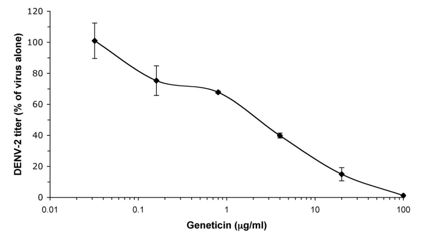
Figure 3 The Geneticin treatment reduces the viral titer/ yield for the DNV2 in a dose dependent manner
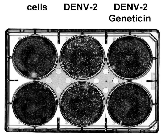
Figure 4 The Geneticin treatment reduces the plaque formation in the dengue virus Type 2 (DENV2) infected cells
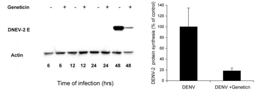
Figure 5 The Geneticin treatment reduces the synthesis of DNEV2 E protein in DNEV infected cells. The reduced width of the protein band in a western blot (in left) and the reduced concentration of synthesized protein in a graph (in right)
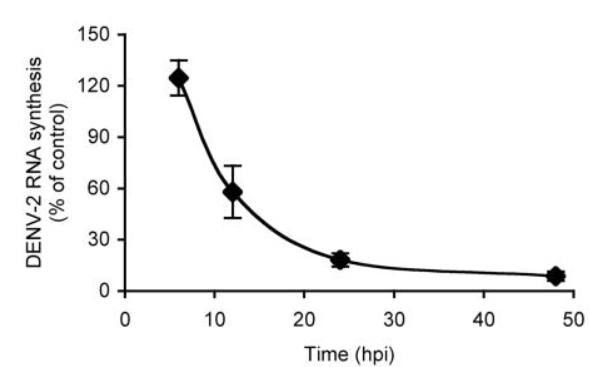
Figure 6 The Geneticin treatment reduces the synthesis of viral RNA as a function of time
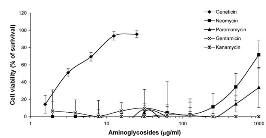
Figure 7 Geneticin is a specific inhibitor of dengue virus Type 1 (DENV2) proliferation, as compared to structurally similar pharmaceutical compounds namely Neomycin, Paromomycin, Gentamicin and Kanamycin. All these compounds despite having a very similar molecular structure to Geneticin could not inhibit dengue virus Type 1 (DENV2) proliferation
More importantly, 2-deoxystreptamine (2-DOS) ring is the main structural feature of Geneticin that lies in the center of the Geneticin molecule. Generally speaking, those Aminoglycosides having 2-deoxystreptamine (2-DOS) ring exhibits the antibiotic properties especially against the bacteria by inhibiting the overall protein synthesis in them by interacting with both subunits of ribosomes; specifically, three main phases, i.e., elongation, termination and recycling, of the translation step are targeted as the movement between the ribosomal subunits is interrupted. These phases are effectively targeted via the interaction of the 2-deoxystreptamine (2-DOS) ring with the secondary structure of the 16S rRNA, i.e., specifically with the major groove of the loop that is evolutionary conserved and asymmetric within the helix 44 (h44) of 16S rRNA. This site is the same that acts as a decoding center in the translation step. This observation is also found in the case of eukaryotic cells. Considering the whole 80S ribosome of the eukaryotic cell, there exist a number of binding sites even for a single type of molecule; for example, for Geneticin alone, there exist four distinct binding sites where four molecules of Geneticin can dock simultaneously. Three of these binding sites are present on the 40S ribosomal subunit which are shown in Figure 8 while the remaining one binding site is present on the 60S ribosomal subunit which is shown in Figure 9 .
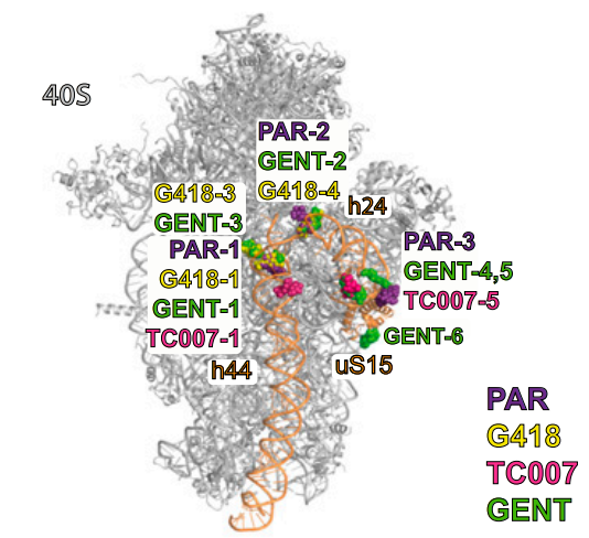
Figure 8 Three Geneticin molecules (shown in yellow and labelled as G418-1, G418-3 and G418-4) are docked into three distinct binding sites present on the 40S ribosomal subunit (smaller subunit) of 80S eukaryotic ribosome
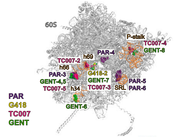
Figure 9 A Geneticin Molecule (shown in yellow and labelled as G418-2) is docked into a distinct binding site present on the 60S ribosomal subunit (bigger subunit) of 80S eukaryotic ribosome
Prion Infection is a landmark of a number of neurogenerative diseases, i.e., diseases that are marked by non-functional nerve cells/ neurons that eventually starts to die, affecting not only human but also non-human species. On the molecular level, this is an infection that is caused by the particles having proteinaceous nature; more appropriately, these particles are called as Prions. Under the normal physiological condition, a protein that is named as prion protein (PrP) is expressed by the host and resides while attached to the outer leaflet of the plasma membrane of the cells of the nervous system particularly nerve cells/ neurons via the glycosylphosphatidylinositol (GPI) anchor. The prion protein (PrP) plays a number of roles with the body but there are two major roles of it; first, to maintain the covering of the myelin sheath in the peripheral nervous system and second, to regulate the neural cell adhesion molecule (NCAM) polysialylation. Under the abnormal pathological condition, the same protein molecules start getting rearranged and changes its native conformation; from alpha helical conformation, it changes to the beta sheets. Resultantly, these altered protein molecules, now termed as PrPSc or PrPres, being resistant to the catalytic activity of the proteases cause neurotoxicity and spread the infection. It has been scientifically shown that the Geneticin treatment upregulates the protein expression of PrP which is the protein in normal conformation (as shown in the western blots in Figure 10), while on the other hand it also downregulates the protein expression of the PrPSc or PrPres which is the protein in abnormal conformation (as shown in the western blots in Figure 11 and graphically in Figure 12). Similar findings have been observed in other cell lines having the prion infection, i.e., CAD5(pIRESneo3), N2a(pIRESneo3), N2a(pcDNA3), CAD5(pIRESneo3), CAD5-PrP−/− (pIRESneo3.HaPrP) and CAD5-PrP−/− (pIRESneo3.HaPrP) as well (data not shown) .
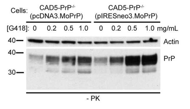
Figure 10 The Geneticin treatment increases the protein expression of the mouse Prion Protein (PrP) in normal configuration in two different cell lines, i.e., CAD5-PrP−/− (pcDNA3.MoPrP) and CAD5-PrP−/− (pIRESneo3.MoPrP) infected with RML prions, in a dose-dependent manner
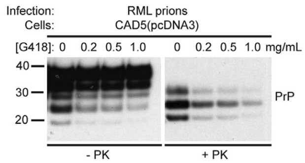
Figure 11 The Geneticin treatment decreases the protein expression of the mouse Prion Protein (PrPres) in abnormal configuration in a cell line, i.e., CAD5 (pcDNA3) infected with RML prions, in a dose-dependent manner as shown in right western blot.
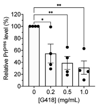
Figure 12 The Geneticin treatment decreases the protein expression of the mouse Prion Protein (PrPres) in abnormal configuration in a cell line, i.e., CAD5 (pcDNA3) infected with RML prions, in a dose-dependent manner
The RNA sequence (1 to 570 nucleotides) of Hepatitis C Virus (HCV), schematically represented in Figure 13, exists in the open conformation as well as in the close conformation as there exists an equilibrium between these two conformations shown in Figure 14; different products are generated from it as shown in Figure 15. The Geneticin treatment inhibits the catalytic activity of RNase Ⅲ and thereby the HCV proliferation is inhibited .

Figure 13 The Schematic representation of the RNA sequence (1 to 570 nucleotides) of Hepatitis C Virus (HCV)
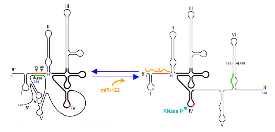
Figure 14 The close conformation (in left) and the open conformation (in right) of the 1 to 570 nt RNA sequence of the Hepatitis C Virus (HCV). In addition, small black arrows indicate sites for RNase Ⅲ while blue one indicates site for RNase P

Figure 15 The products after the activity of RNase Ⅲ




Comentarios