Detection principle
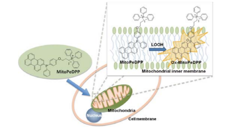
MitoPeDPP can cross cell membranes and accumulate in mitochondria. MitoPeDPP accumulated on the inner mitochondrial membrane can be oxidized by lipid peroxides and release strong fluorescence.
1. Example of co-staining with MitoPeDPP and mitochondrial staining reagent MitoBright
Adding t-BHP (tert-butyl hydroperoxide) to HeLa cells to detect lipid peroxides
Wavelength (wavelength/band pass)
MitoPeDPP: 470/40 (Ex), 525/50 (Em)
MitoBright DeepRed: 600/50 (Ex), 685/50 (Em)
The results confirmed that MitoPeDPP fluoresces after being oxidized by t-BHP in the mitochondria of HeLa cells. In addition, through co-staining with mitochondrial staining reagent (MitoBright Deep Red: MT08), it was confirmed that the fluorescence of MitoPeDPP is localized in mitochondria.

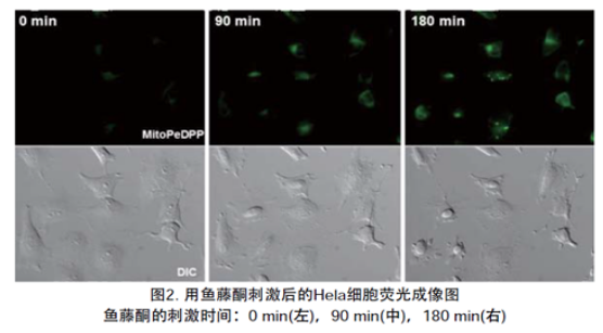
2. Detect lipid peroxides produced by adding Rotenone
After adding MitoPeDPP to HeLa cells [μ-slide, 8-well (manufactured by Ibidi)], add Rotenone solution and observe using a fluorescence microscope. Experimental results confirmed that after adding Rotenone, the production of lipid peroxides in cells was detected.
Rotenone stimulation time: 0 min (left), 90 min (middle), 180 min (right)
3.Experimental example of using MitoPeDPP on nerve cells
A. Fluorescence microscopy detection
Isoflavin was added to NIE-115 cells (mouse neuroblastoma) to induce Ca2+ influx into the cells, and the production of lipid-soluble peroxides in the mitochondrial membrane was observed by fluorescent staining with MitoPeDPP. The experimental results confirmed that the experimental group added with isomycin had stronger fluorescence than the control group.
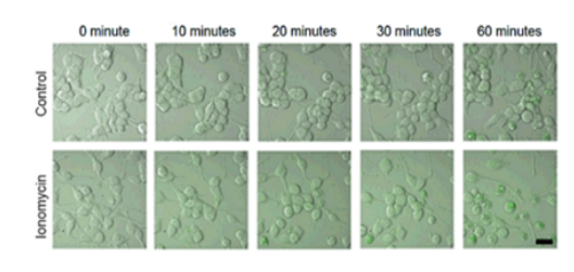
B. Comparison of average fluorescence intensity data
To quantify the fluorescence intensity of control cells and cells with added ionomycin, a comparison of the two sets of data was performed based on the mean fluorescence intensity.
The results confirmed that the fluorescence intensity observed in the cells 30 minutes after adding ionomycin was significantly increased compared to the cells in the control group.
Data provided by Free Radical Research, in press
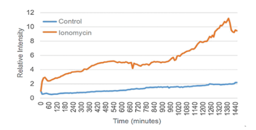
Refer to Shibaura Institute of Technology Institute of Technology Associate Professor Fukui Koji and Nakamura Saki [Reference 3]
4. Selectivity of MitoPeDPP reaction
In cell-free reaction systems, MitoPeDPP can react with various peroxides such as H2O2, t-BHP and ONOO-, but in cells, accumulation
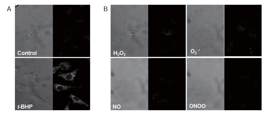
MitoPeDPP accumulated in mitochondria can be oxidized by t-BHP to release strong fluorescence (Figure 3A), but reacts only weakly with other ROS or RNS (Figure 3B).
A) MitoPeDPP was added to HepG2 cells and cultured for 15 min, then treated with 100 μmol/l t-BHP.
B) Add MitoPeDPP to HepG2 cells and culture for 15 minutes, then add ROS and RNS inducers.
Add 100 μmol/l (H2O2, NO and ONOO-inducer) and 10 μmol/l PMA (O2-.inducer) respectively.
The left side is the bright field image and the right side is the fluorescence image.
* t-BHP: tert-Butylhydroperoxide; PMA, Phorbol myristate acetate;
SIN-1, 3-(Morpholinyl)sydnonimine, hydrochloride;
NOC 7, 1-Hydroxy-2-oxo-3-(N-methyl-3-aminopropyl)-3-methyl-1-triazene
Wavelength/bandpass filter: 470/40 (Ex), 525 /50 (Em)
References:
1) K. Shioji K, Y. Oyama, K. Okuma and H. Nakagawa, "Synthesis and properties of fluorescence probe for detection of peroxides in mitochondria.", Bioorg Med Chem Lett., 2010, 20, (13), 3911.
2) S. Oka, J. Leon, K. Sakumi, T. Ide, D. Kang, F. M. LaFerla and Y. Nakabeppu, "Human mitochondrial transcriptional factor A breaks the mitochondria-mediated vicious cycle in Alzheimer’s disease", Scientific Reports ., 2016, DOI: 10.1038/srep37889 , .
3) S. Nakamura, A. Nakanishi, M. Takazawa, S. Okihiro, S. Urano and K. Fukui, "Ionomycin-induced calcium influx induces neurite degeneration in mouse neuroblastoma cells: Analysis of a time-lapse live cell imaging system", Free Radical Research., 2016, 50, (11), 1214.
4) M. Akimoto, R. Maruyama, Y. Kawabata, Y. Tajima and K. Takenaga, "Antidiabetic adiponectin receptor agonist AdipoRon suppresses tumour growth of pancreatic cancer by inducing RIPK1/ERKdependent necroptosis", Cell Death Dis., 2018, 9, 804.
5) M. Álvarez-Córdoba, A. Fernández Khoury, M. Villanueva-Paz, C. Gómez-Navarro, I. Villalón-García, J. M. Suárez-Rivero, S. Povea-Cabello, M. Mata, D. Cotán, M. Talaverón-Rey, A. J. Pérez-Pulido, J. J. Salas, E. M. Pérez-Villegas, A. Díaz-Quintana, J. A. Armengol, J. A. Sánchez-Alcázar , "Pantothenate Rescues Iron Accumulation in Pantothenate Kinase-Associated Neurodegeneration Depending on the Type of Mutation.", Mol. Neurobiol. ., 2019, 56, (5), 3638.
6)Yaxu Li, Qiao Ran, Qiuhui Duan, Jiali Jin, Yanjin Wang, Lei Yu, Chaojie Wang, Zhenyun Zhu, Xin Chen, Linjun Weng, Zan Li, Jia Wang, Qi Wu, Hui Wang, Hongling Tian, Sihui Song, Zezhi Shan, Qiwei Zhai, Huanlong Qin, Shili Chen, Lan Fang, Huiyong Yin, Hu Zho“7-Dehydrocholesterol dictates ferroptosis sensitivity”Nature.626, 411-418(2024).


