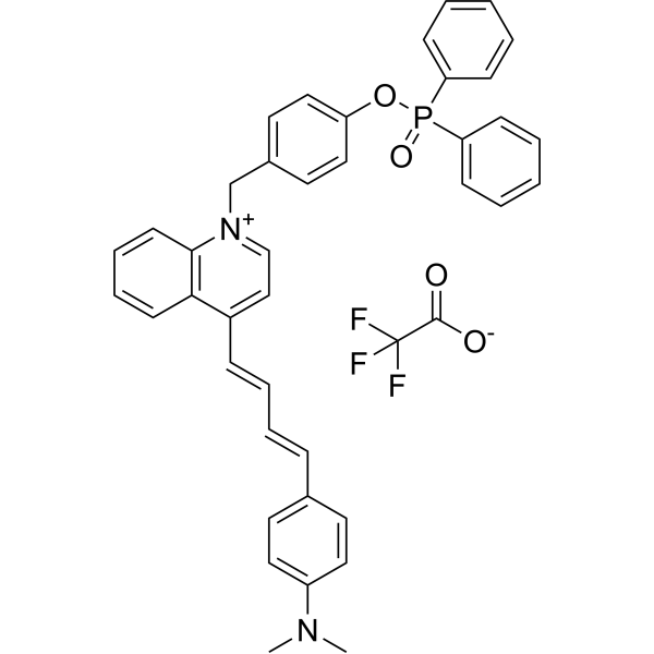
MQA-P TFA |
| Catalog No.GC71077 |
MQA-P TFA es una sonda fluorescmultifuncional en infrarrojo cercano (NIR) que detecta simultáneamente ONOO-, viscoy polaridad dentro de las mitocondrias.
Products are for research use only. Not for human use. We do not sell to patients.

Sample solution is provided at 25 µL, 10mM.
Guidelines (Following is our recommended protocol. This protocol only provides a guideline, and should be modified according to your specific needs).
1. MQA-P is dissolved in dimethyl sulfoxide (DMSO) to prepare a stock solution (1.0 mM).
2. For imaging of ONOO- in live cells.
HeLa cells are incubated with MQA-P (5 μM) for 30 min as control; pretreated with SIN-1 for 30 min and then incubated with MQA-P (5 μM) for another 30 min. The fluorescence images are obtained on a confocal laser scanning microscope with a green channel (λex= 405nm, λem= 550-670 nm).
3. For imaging of viscosity in live cells.
HeLa cells were incubated with MQA-P (5 μM) for 30 min as control; pretreated with Monensin for 30 min and then incubated with MQA-P (5 μM) for another 30min. The fluorescence images are obtained on a confocal laser scanning microscope with a red channel (λex= 561 nm, λem= 680-750 nm).
4. For dual-channel imaging of ONOO-, viscosity and polarity during ferroptosis.
HeLa cells are incubated with MQA-P (5 μM) for 30 min as control; pretreated with Erastin for 30 min and then incubated with MQA-P (5 μM) for another 30 min. The fluorescence images are obtained on a confocal laser scanning microscope with a green channel (λex= 405nm, λem= 550-670 nm) for ONOO- and a red channel (λex= 561 nm, λem= 680-750 nm) for viscosity and polarity[1].
Guidelines (Following is our recommended protocol. This protocol only provides a guideline, and should be modified according to your specific needs).
1. For tissue slices imaging, the normal organs (including heart, liver, spleen, lung, and kidney) and tumor are isolated from the mice, then sectioned as 5 μm thicknesses, respectively.
2. These slices are incubated with MQA-P (20 μM) for 30 min, then washed with PBS (pH 7.4) three times, and finally subjected to in vivo imaging using a confocal laser scanning microscope with a green channel (λex=405nm, λem=550-670 nm) for ONOO- and a red channel(λex=561 nm, λem=680-750 nm) for viscosity and polarity, respectively[1].
References:
[1]. Li Fan, et al. Multifunctional Fluorescent Probe for Simultaneous Detection of ONOO-, Viscosity, and Polarity and Its Application in Ferroptosis and Cancer Models. Anal Chem. 2023 Apr 4;95(13):5780-5787.
Average Rating: 5 (Based on Reviews and 30 reference(s) in Google Scholar.)
GLPBIO products are for RESEARCH USE ONLY. Please make sure your review or question is research based.
Required fields are marked with *


